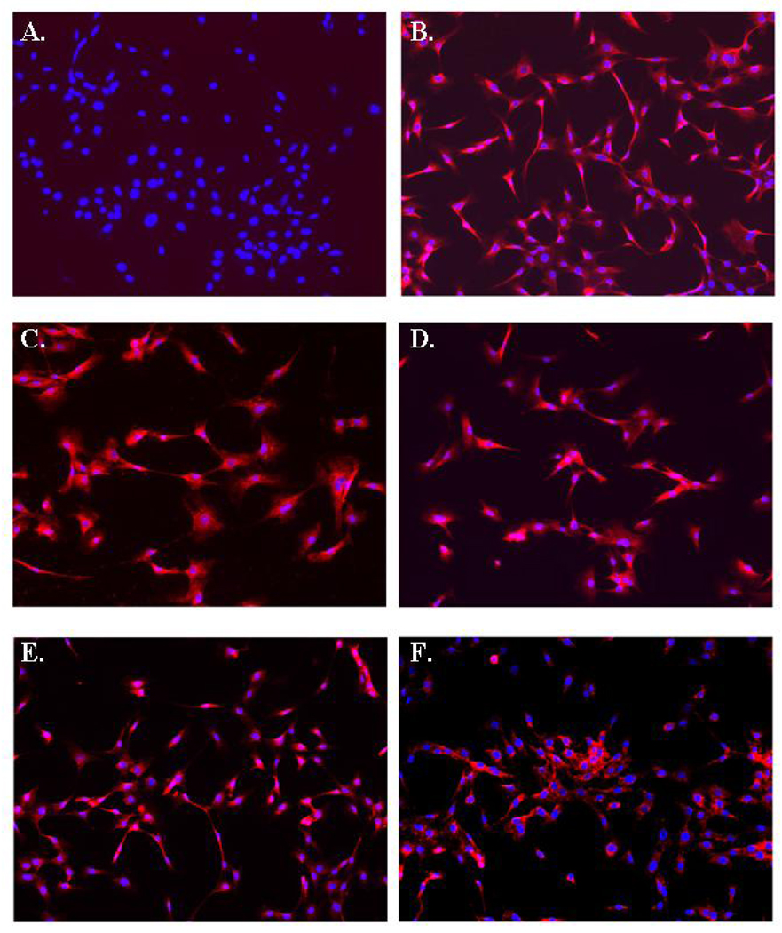
Figure 2.
Immunohistochemical analyses of GFAP, Glutamate Receptor 1, NMDA Receptor, EAAT1, and EAAT2 on astrocytes isolated from H-2Kb-tsA58 mice. (A) Control labeling with secondary antibody only. (B) All astrocytes stain positive for GFAP expression. (C) Homogenous staining was observed for glutamate receptor 1 and (D) NMDA receptor. (E) EAAT1 and (F) EAAT2 expression on cultured astrocytes. Original magnification × 200.

