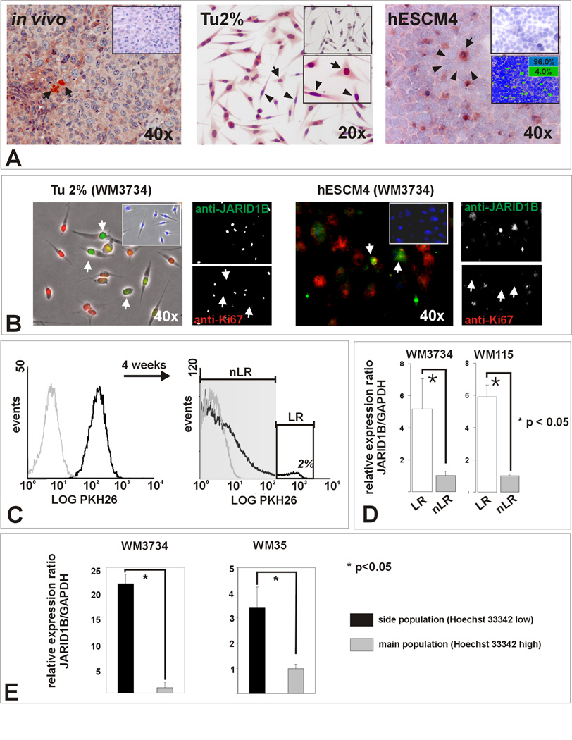
Figure 1. Melanomas contain a subpopulation of slow-cycling cells characterized by increased JARID1B expression.
(A) Left panel: Representative example of a vertical growth phase melanoma after immunostaining for JARID1B. Single positive cells (arrows) with predominantly nuclear and minor cytoplasmic staining are surrounded by negative cells. Middle: Single adherent WM3734 melanoma cells show high JARID1B (arrows) while the bulk of cells shows median or low expression (arrowheads) when grown in conventional medium (Tu2%). Isotype control (top) and higher magnification (bottom) are shown as inserts. Right: More distinct JARID1B staining was seen in cryosections of WM3734 melanoma spheres (hESCM4). Digital quantitation after pseudocoloring revealed a relative frequency of 4% JARID1B-immunopositive (green) and 96% immunonegative cells (blue) for this section (see also Figure S1). (B) Anti- JARID1B-positive cells (Alexa Fluor® 488, green) are predominately negative for the proliferation marker Ki-67 (Alexa Fluor® 568, red). The result was reproduced in WM115 cells (not shown). (C) Flow cytometry analysis of WM3734 melanoma sphere cells after incubation with PKH26 (black line) compared to unlabeled control cells (grey line). After 4 weeks in hESCM4, 2% of the cells retained the maximum label (label-retaining ‘LR’ cells, white square). (D) LR cells displayed significantly enhanced JARID1B expression compared to non label-retaining (nLR) cells in QPCR. (E) QPCR screening of side population cells from WM3734 and WM35 melanoma sphere cells showing an upregulation of JARID1B compared to the main populations.

