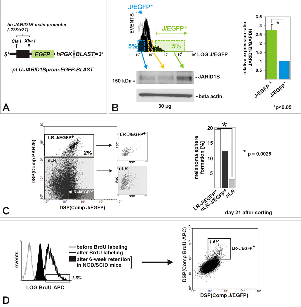
Figure 2. JARID1B is a biomarker for the label-retaining subpopulation in melanoma.
(A) Lentiviral pLU-JARID1Bprom-EGFP-Blast. (B) Flow cytometric determination of the J/EGFP-positive subpopulation, exemplarily shown for WM3734JARID1Bprom-EGFP melanoma spheres grown in hESCM4 (see also Figure S2). QPCR and immunoblots of sorted populations showed a significant correlation between exogenous EGFP and endogenous JARID1B expression (p<0.05, t-test). The 5%-threshold was based on our in vitro and in vivo observations on endogenous JARID1B expression frequency (Figure 1). (C) PKH26 label-retention of WM3734JARID1Bprom-EGFP sphere cells in vitro. Left: LR cells grouped as a distinct J/EGFP-positive subpopulation (LR/JEGFP-positive, white square) and were enriched for small-sized cells. Right: LR-J/EGFP-positive cells (white bar) showed the highest capacity to re-form spheres. Depicted is one representative out of three independent experiments. (D) Left: Anti-BrdU-APC flow cytometry of dissociated tumor cells revealed that 6 weeks after BrdU incubation the majority of cells diluted out the BrdU (filled black). Unlabeled (grey line) and freshly BrdU-labeled cells (black line) served as controls. The threshold was set to the peak APC intensity of freshly BrdU-labeled cells. Right: In vivo BrdU-labeled cells grouped as a distinct J/EGFP-positive subpopulation (LR/JEGFP-positive, white square, 1.6% of total population).

