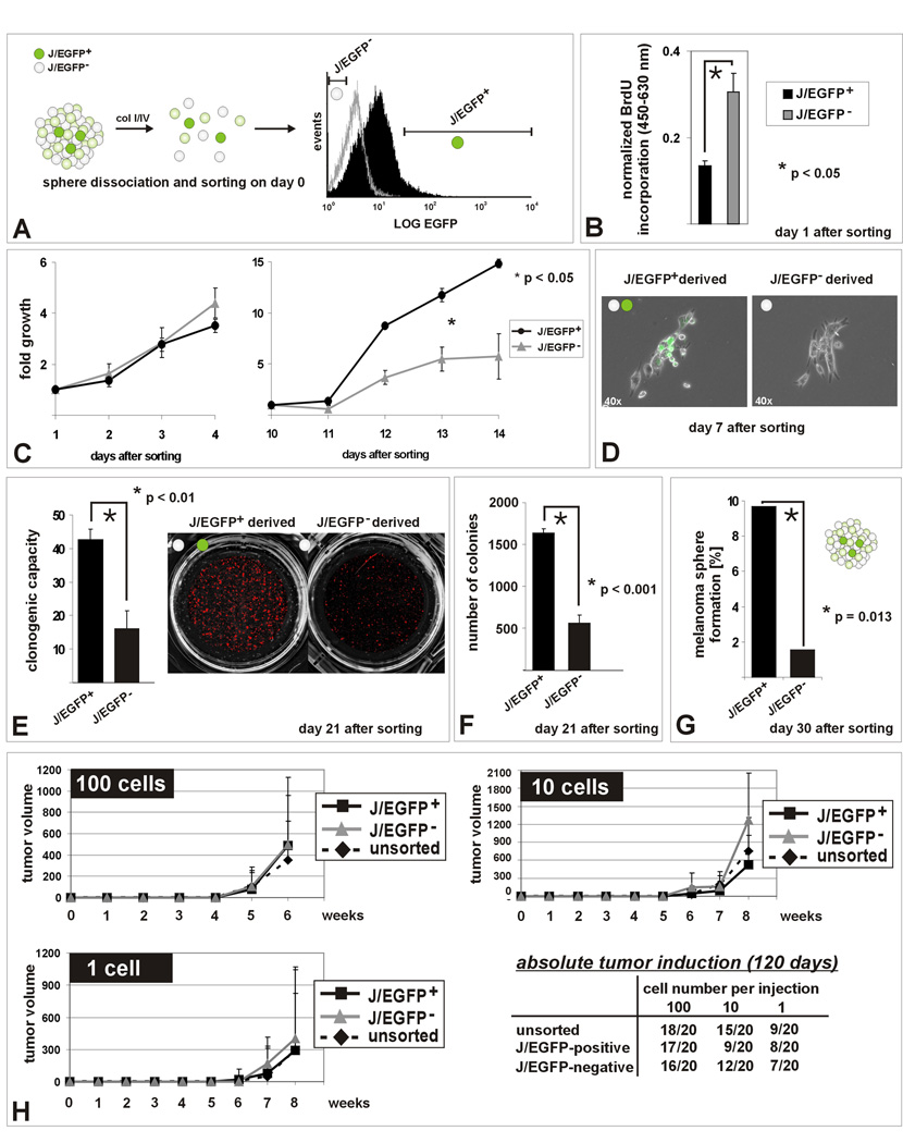
Figure 3. In vitro self renewal capacity of the JARID1B-positive subpopulation.
(A) Methodical scheme. (B) Decreased BrdU incorporation into J/EGFP-positive cells 24 hours after separation from spheres (hESCM4) as determined by ELISA after pulsed BrdU exposition. (C) MTS assays of sorted J/EGFP-positive and –negative cells in hESCM4 (see also Figure S3). After day 10, the progeny of J/EGFP-positive cells proliferated significantly faster and (D) microscopically consisted of J/EGFP-positive and an increasing number of J/EGFP-negative cells. (E) Enhanced clonogenicity of single J/EGFP-positive cells. (F) J/EGFP-positive cells had both a higher potential to form 3D-colonies in 0.35% hESCM4-soft agar and (G) to self-renew again into heterogeneous melanoma spheres in limited dilution assays (hESCM4). Shown are representative results from at least two independent experiments. (H) Xenotransplantation growth curves after subcutaneous injection of 100, 10 or 1 WM3734JARID1Bprom-EGFP melanoma cell into NOD/LtSscidIL2Rγnull mice. Growth curves were stopped when the first mouse of the respective series exceeded a tumor size >1000 mm3. Also during the subsequent observation of remaining mice, no differences were seen.

