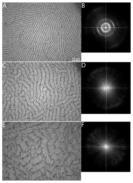
Figure 2.

Representative experimental microscope images of self-assembled Au nanowires with nominal lengths of 2 (a), 4 (c), and 7 μm (e). The corresponding FTs of the images in a, c, and e (b, d, and f, respectively) are included to the right of the corresponding microscope image. The FTs were cropped to magnify the features in the center. The scale bar in (a) is 10 μm and applies to images (c) and (e).
