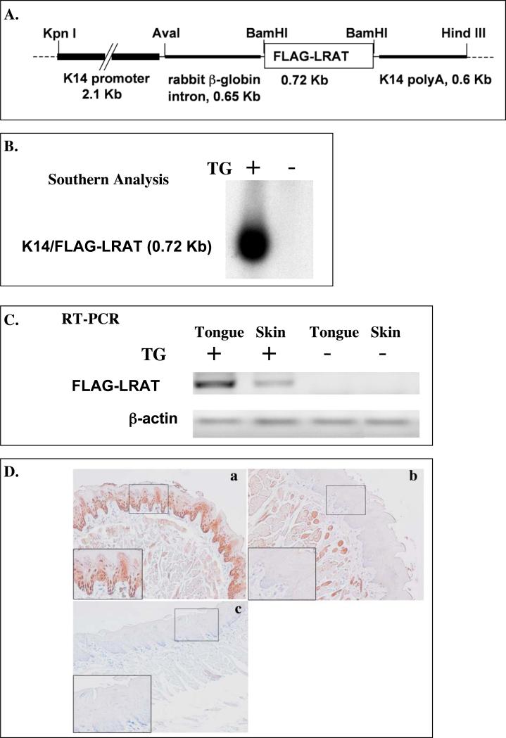
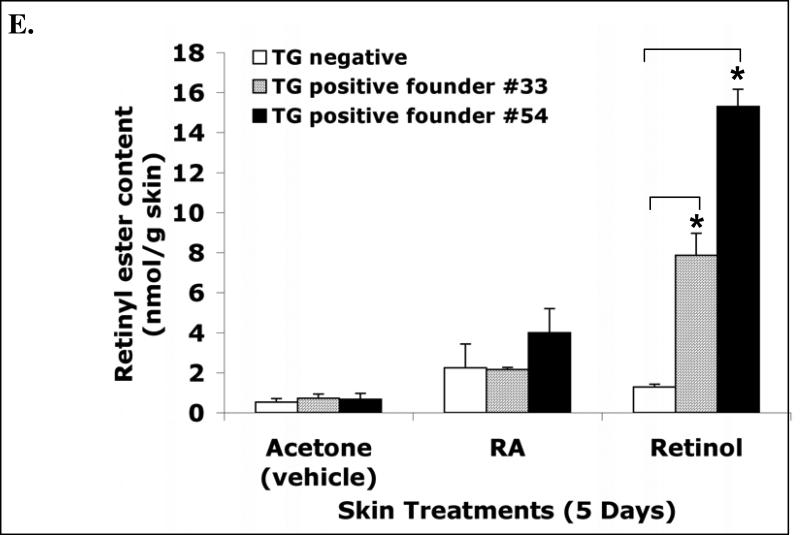
Figure 1. Targeted overexpression of the transgene FLAG-hLRAT to the mouse epithelial basal layer cells.
A, The schematic structure of the transgene cassette. B, Southern blot result shows the presence of the transgene in the mouse genomic DNA. C, RT-PCR analysis of the FLAG-hLRAT transgene mRNA in the tongue and skin of K14/FLAG-hLRAT Southern blot transgenic positive (TG+) and negative (TG-) mice from founder #54. D, The expression of the transgene human LRAT protein in the tongue epithelial basal layer cells of K14/FLAG-hLRAT transgenic positive (TG+) and negative (TG-) mice from founder #54 (200 ×). K14/FLAG-hLRAT TG+ and TG- mice were sacrificed, and the tongues were fixed, embedded, sectioned, and stained with anti-human LRAT antibody (see “Materials and Methods”). a, TG positive mouse tongue; b, TG negative mouse tongue; c, negative control, tongue stained only with a secondary antibody; the inset in each picture is 3 × the digital magnification of the small boxed area in the picture. E, the levels of retinyl esters in the skin of K14/FLAG-hLRAT TG positive and negative mice examined by HPLC after a 5 day topical treatment with 40 nmoles (volume 400 μl) of all-trans retinoic acid (ATRA) or retinol per day. Drugs were dissolved in acetone that was the vehicle (see “Materials and Methods”). TG+ #33 and #54 are two TG positive founder lines. The differences among different treatment groups were analyzed by using a one way ANOVA test. Differences with a p value of < 0.05 (marked with an asterisk) in a comparison of retinyl ester levels in retinol treated TG+ mouse skin to that in retinol treated TG- mouse skin were considered to be statistically significant.


