
Figure 3.
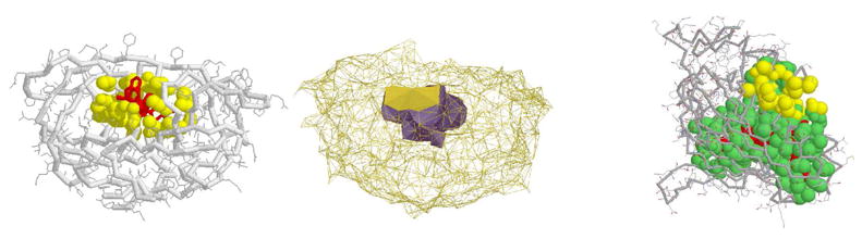
The binding pockets on HIV-1 protease and phosphatidylinositol transfer protein (PITP). (Left): Binding pocket (yellow) on HIV-1 shown in van der Waals space filling model. Ligand is colored red. (Middle): The alpha shape of the HIV-1 binding site. Its mouth opening is colored gold. (Right): Binding pocket (green) on PITP for phoshpolipid (red) and a regulatory site on a different region (yellow) of the same protein.