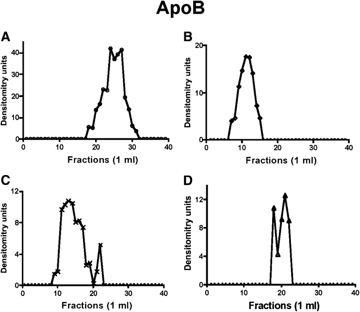
Fig. 1.
Distribution of ER-apoB48 across a Sephacryl S-400 HR column under differing conditions. In each case (A–D), ER membranes (1 mg protein) were treated as indicated, collected by centrifugation, washed with PBS, and solubilized in 1% Triton X-100. The ER protein was chromatographed over a 1% Triton X-100 equilibrated Sephacryl S-400 HR column (see “Methods”), and 1-ml fractions were collected. Each fraction was tested for apoB48 by immunoblot. The band densities were quantitated by the GelDoc XR imaging system. The resulting arbitrary density units are reported. (A) Native ER membranes. (B) ER incubated with native cytosol and ATP. (C) ER membranes incubated with rL-FABP without cytosol. (D) Urea-washed ER incubated with PKCζ immunodepleted cytosol and ATP (see “Methods”). In A–C, incubations were at 4°C; in D, the incubation was at 37°C (see “Methods”). Apo, apolipoprotein; ER, endoplasmic reticulum; L-FABP, liver fatty acid-binding protein; PKC, protein kinase C.

