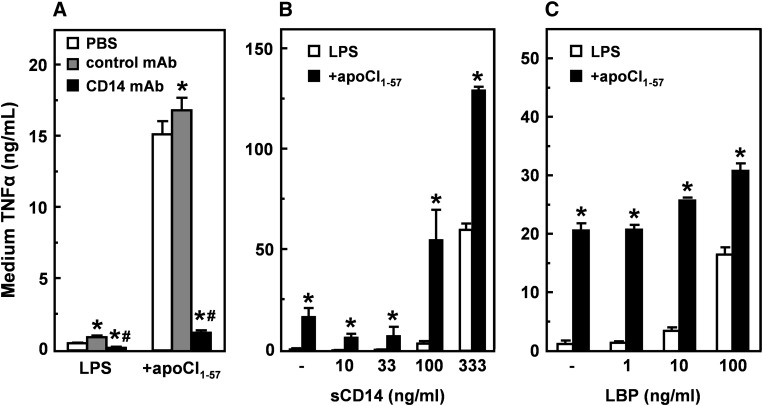
Fig. 8.
Role of CD14 and LBP in the stimulating effect of apoCI on the LPS-induced TNFα response in vitro. A: RAW 264.7 cells were preincubated (30 min at 37°C) in DMEM supplemented with 0.01% human serum albumin and vehicle (white bars), isotype control antibody (gray bars), or anti-CD14 antibody (black bars), washed, and subsequently incubated (4 h at 37°C), in DMEM supplemented with 0.01% human serum albumin, with LPS (1 ng/ml) that was preincubated (30 min at 37°C) without or with a 100-fold molar excess of apoCI1–57. TNFα was determined in the medium by ELISA. B, C: RAW 264.7 cells were incubated (4 h at 37°C), in DMEM supplemented with 0.01% human serum albumin, with LPS (1 ng/ml) that was preincubated (30 min at 37°C) without or with a 100-fold molar excess of apoCI1–57 in the presence of soluble CD14 (sCD14) (B) or LBP (C). TNFα was determined in the medium by ELISA. Data are expressed as mean TNFα concentration ± SD (n = 3–4). *P < 0.05 compared with vehicle (A) or LPS alone (B, C); #P < 0.05 compared with control antibody.

