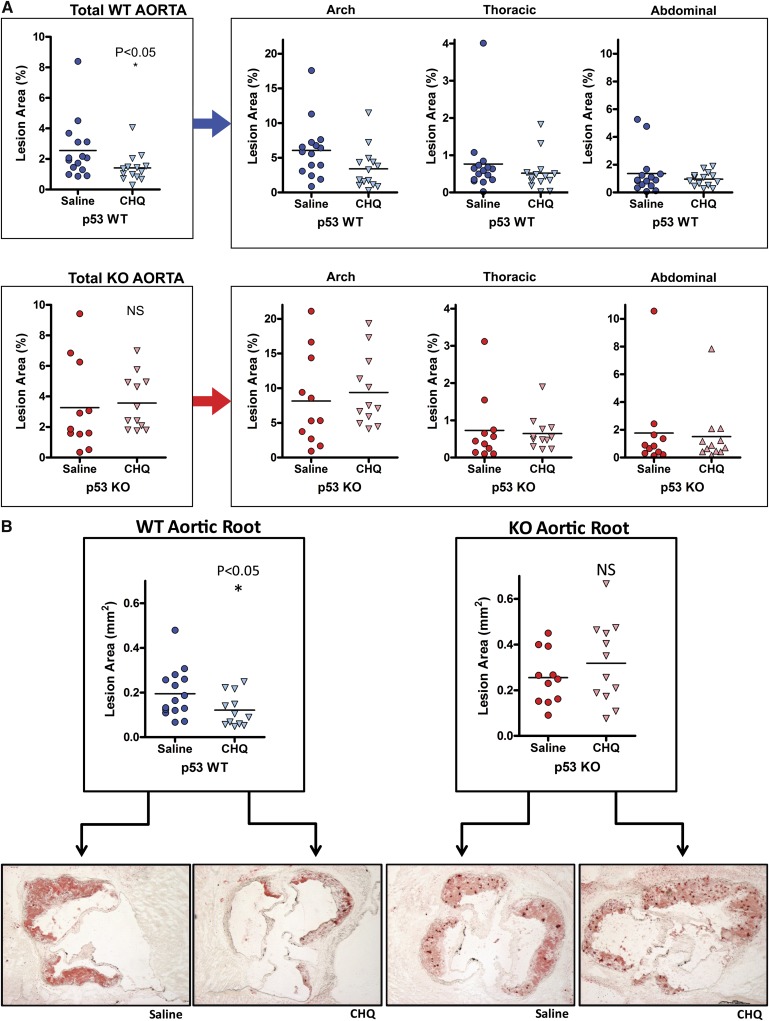
Fig. 5.
Effects of chloroquine on atherosclerosis in the presence and absence of p53. Atherosclerotic area involvement was determined by computer image analysis of (A) en face pinned aortas for the total aorta (p53 WT, upper-left panel; p53 KO, lower-left panel), and regions of the aorta (aortic arch, thoracic aorta, and abdominal aorta, right panels), and (B) Oil-Red-O–stained sections of the aortic roots (p53 WT, left panel; p53 KO, right panel). Representative aortic root sections are shown below the data sets. Horizontal lines within the data sets represent medians. *P < 0.05. CHQ, chloroquine; KO, knockout; NS, not significant; WT, wild type.

