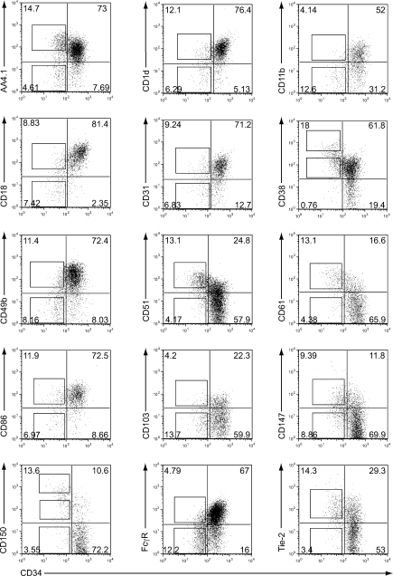
Figure 1.
Markers with heterogeneous expression in CD34−KSL cells. KSL cells were stained with FITC-conjugated anti-CD34 antibody and additional PE-conjugated antibodies as shown. Shown are flow cytometric profiles for the markers that had heterogeneous expression within CD34−KSL cells (percentages are shown). Marker-positive or -negative cells were separated by using the sorting gates (shown as squares). In the case of CD150, CD34−KSL cells were separated into CD150high, CD150med, and CD150neg fractions. In the case of CD38, CD34−KSL cells were separated into CD38high and CD38med fractions. The data represent four to eight independent experiments.

