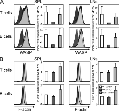
Figure 2.
WASP-I296T and WASP-L272P are expressed in B and T cells and induce marked increase in polymerized actin. (A) WASP expression. Spleen and lymph node T and B cells were stained for WASP using an anti-WASP antibody followed by flow cytometric analysis. (B) Polymerized actin (F-actin) content. Spleen and lymph node T and B cells were stained with phalloidin to detect F-actin and analyzed by flow cytometry. Each panel shows one representative histogram (left) and one graph (right) of mean values (±SD) of six experiments (n = 6; A) and five experiments (n = 5; B).

