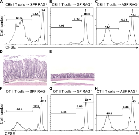
Figure 3.
CD4+ T cells do not undergo spontaneous proliferation in germfree RAG−/− mice. 106 CFSE-CBir1 Tg CD4+ T cells (A–C) or 106 CFSE-OT II CD4+ T cells (F–H) were transferred into RAG−/− mice housed under SPF conditions (A and F), germfree (GF) conditions (B and G), or ASF conditions (C and H). 10 d after the transfer, division of the CFSE-labeled CD4+ T cells was determined by CFSE dilution. Numbers in each plot represent the percentage of donor cells undergoing spontaneous proliferation, homeostatic proliferation, and staying undivided, respectively. Data are representative of at least three individual mice of each group from three independent experiments. Histopathology of the colon of CBir1 Tg T cell recipients at 5 mice/group under SPF conditions (D) and germfree conditions (E) was assessed 8 wk after cell transfer. Bars, 100 µm. Data are representative of three independent experiments with similar results.

