Figure 3.
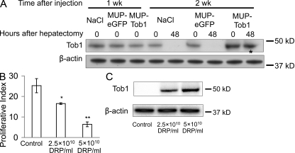
MUP-Tob1-AAV8 virus inhibits hepatocyte proliferation after hepatectomy in a dose-dependent fashion. (A) Western blot of mouse liver after injection of MUP-Tob1-AAV8, MUP-eGFP-AAV8, or NaCl. The asterisk denotes Tob1 expression maintained even 48 h after hepatectomy and 2 wk after injections. (B) Tob1-null mice were infected with indicated doses of MUP-Tob1-AAV8 virus or MUP-eGFP-AAV8 (control). 3 wk later, 2/3 hepatectomy was performed and livers analyzed 48 h after hepatectomy. BrdU labeling is shown. n = 3. *, P = 0.01; **, P < 0.05. (C) Western blot of Tob1 in whole cell lysates. β-Actin is used to demonstrate equal protein loading. All blot lanes are the most representative sample from among at least three different animals.

