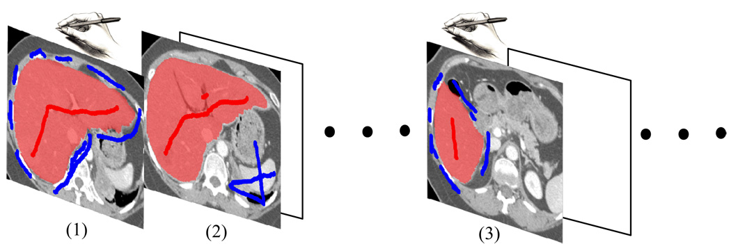
Figure 1.
The process of segmenting a stack of medical images in our method. (1) On one of the images, the user specifies the seed pixels interactively by using brush strokes. At this time no statistics is available for segmentation. Once the initial result is satisfactory, pixels along the boundary are sampled. (2) On the subsequent image slices, the boundary term is estimated from the samples on the training slice. The regional term is also estimated from the new brush strokes on the image slice being segmented. Note that the humane interactions (brush strokes) are significantly reduced. (3) Users can always re-train the model if the boundary statistics is no longer applicable.

