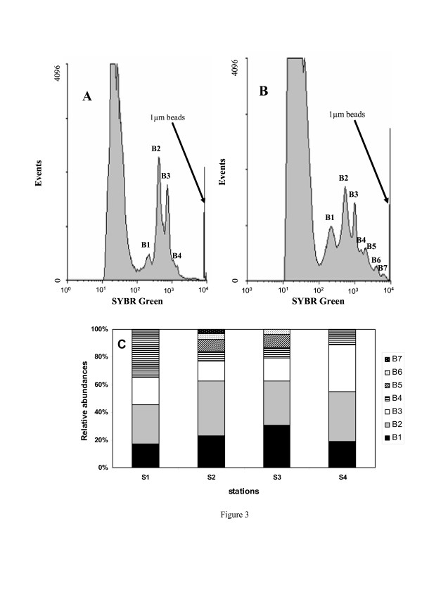
Figure 3.
Cytometric differentiation of prokaryotic populations. (A) Shows results obtained from S4. Four prokaryotic sub-populations were identified; the histogram plot of green fluorescence shows 4 peaks relating to sub-populations of increasing DNA content (B1 to B4). (B) Shows results obtained from S2. Seven prokaryotic sub-populations were identified; the histogram plot of green fluorescence shows 7 peaks relating to sub-populations of increasing DNA content (B1 to B7). Sub-populations differed through their green fluorescence and side scatter, and therefore were not classified into high and low DNA-subpopulations but as different discrete populations. (C) Relative abundances (%) of cytometrically-defined sub-populations along the salinity gradient from S1 to S4.

