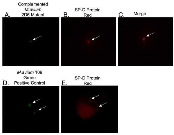
Figure 5.
Fluorescent microscopy images of U937 macrophages infected with fluorescein-labeled complemented M. avium 2D6 mutant. The SP-D protein is shown in red. Arrows point to bacteria (green) and SP-D protein (red). SP-D is present in macrophages infected with the MAC 104 strain and absent in the 2D6 mutant-infected macrophages.

