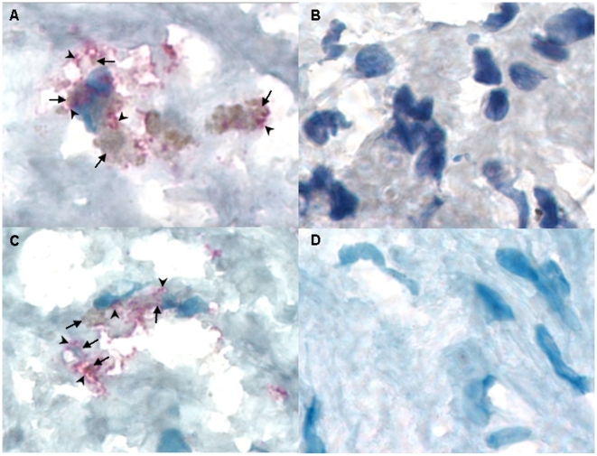
Figure 1. Carotid plaque sections showing co-localization of Lp-PLA2 and C. pneumoniae, and macrophages and C. pneumoniae.
A) Lipoprotein-associated phospholipase A2 (Lp-PLA2) was detected by Immunohistochemistry (IHC) using anti- Lp-PLA2 specific monoclonal antibody (diaDexus) and chromagen fast red (arrowheads); C. pneumoniae was detected by IHC using a CHsp60-specific MAb and chromagen DAB (arrows); ×1000. B) Negative control of a positive carotid plaque section (same patient as in A); ×1000. C) Carotid plaque section showing co-localization of C. pneumoniae (DAB; arrows) and macrophages detected by IHC using CD68 macrophage-specific monoclonal antibody and chromagen fast red (arrowheads); ×1000. D) Negative control of a positive carotid plaque section (same patient as in C).

