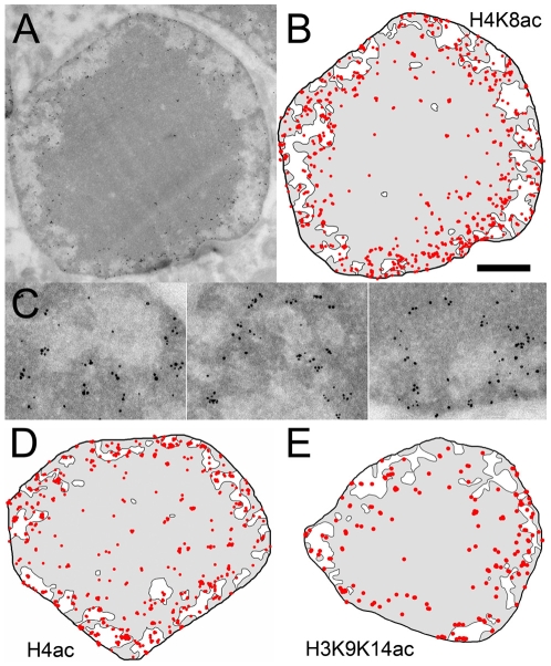
Figure 5. Immunolabelling of acetylated histone marks.
(A) Overall view of a rod nucleus immunogold labelled with acetylated H4K8-specific antibodies. (B) Schematic representation of the H4K8ac-specific labelling. (C) Enlarged region showing the distribution of the H4K8ac labelling around the EC cavities. (D) Schematic representation of the labelling for the pan-actetylated H4. (E) Schematic representation of the labelling for H3 actetylated on K9 and K14. The bar represents 1 µm in A, B, D, E, and 260 nm in C.

