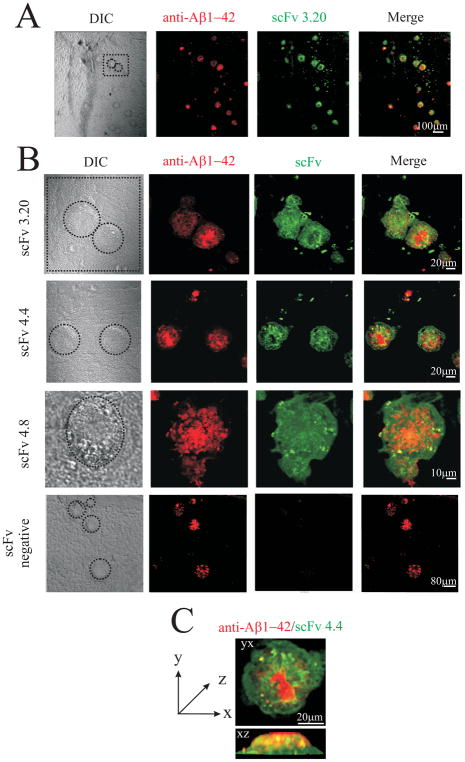
Fig. 6.
Purified soluble scFv antibodies from human non-immune phage display library specifically recognize Aβ deposits in the hippocampus of transgenic mice (Tg2576). Each panel shows, from the left: the reactivity of a rabbit anti-Aβ1-42 antibody (shown in red); the reactivity of soluble scFv fragments (shown in green); the merge between red and green channels. (A) Low magnification provides an overview of the hippocampus in which amyloid deposits are recognized by the soluble scFv fragment (scFv3.20). (B) High magnification provides a detailed view of binding of scFv 3.20, scFv 4.4 and scFv 4.8 to amyloid deposits. Merged images illustrate clear colocalization of red and green fluorescence (in yellow) as well as only-green and only-red areas. Only a background staining is observed with a control scFv antibody. (C) Three-dimensional reconstruction of sequential confocal sections (z-stacks, taken 0.3–0.45 μm apart in stacks) of an amyloid deposit in yx and xz planes.

