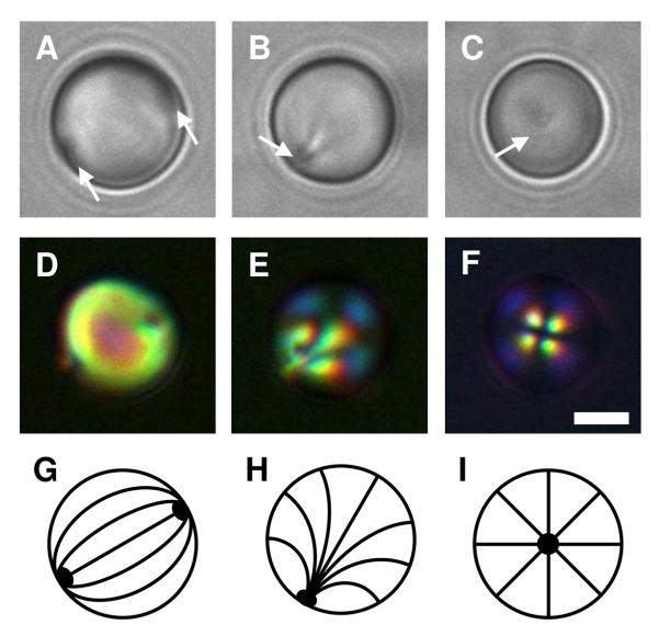
Figure 1.
A-C) Bright-field and D-F) polarized light micrographs of dispersed 5CB droplets incubated in A, D) a buffer solution (10 mM HEPES, pH 7); B, E) a solution of polymer 1 at 0.1 mg/mL; and C, F) a solution of polymer 1 at 1.0 mg/mL. The 5CB emulsions were under continuous agitation for 2 h. G-I) Schematic illustrations of the director profiles for G) bipolar, H) preradial, and I) radial configurations. Point defects in the 5CB droplets shown in bright-field images are indicated by the white arrows. Scale bar = 3 μm.

