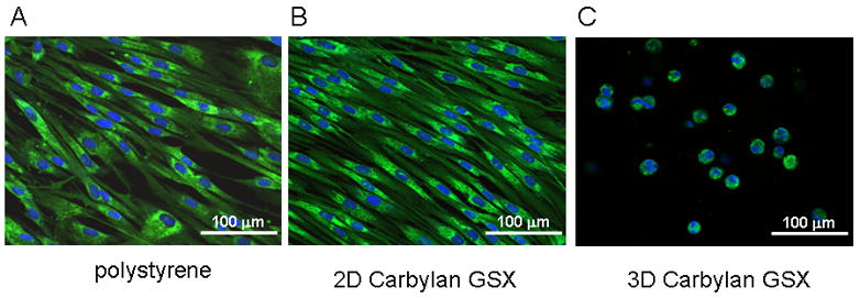
Figure 2.

Morphologic appearance and fibroblast marker (hPH) distribution of immortalized hVFFs in various culture environments. After 72h of in vitro culture, fluorescent images of hVFF on polystyrene (A), on a thin layer of Carbylan GSX (2D condition) or in Carbylan GSX (3D condition) were captured by an inverted confocal microscope equipped with dual excitation lasers for green and blue fluorescence.
