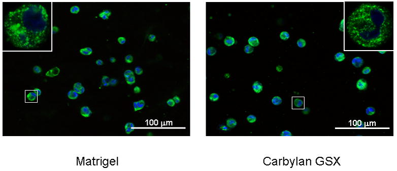
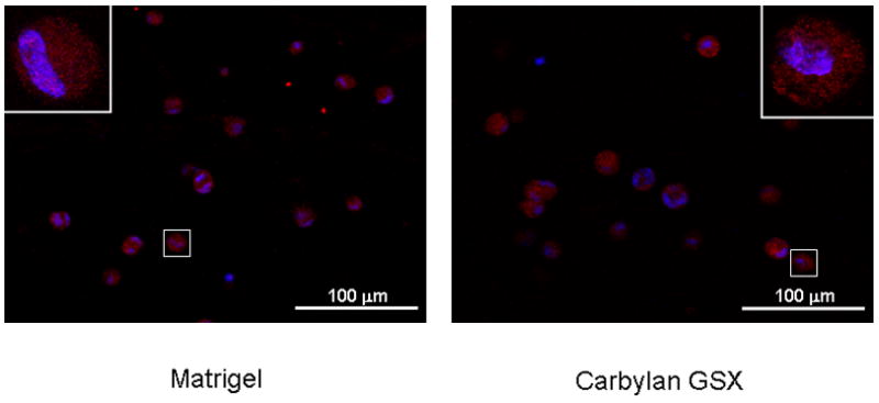
Figure 3.


The distribution of hPH (A) and ICAM-1 (B) in hVFF cultured in 3D Matrigel and Carbylan GSX environments. After 72h of in vitro culture, the fluorescent images of hVFF in Matrigel and Carbylan GSX (3D condition) were captured by an inverted confocal microscope equipped with dual excitation lasers for red, green and blue fluorescence.
