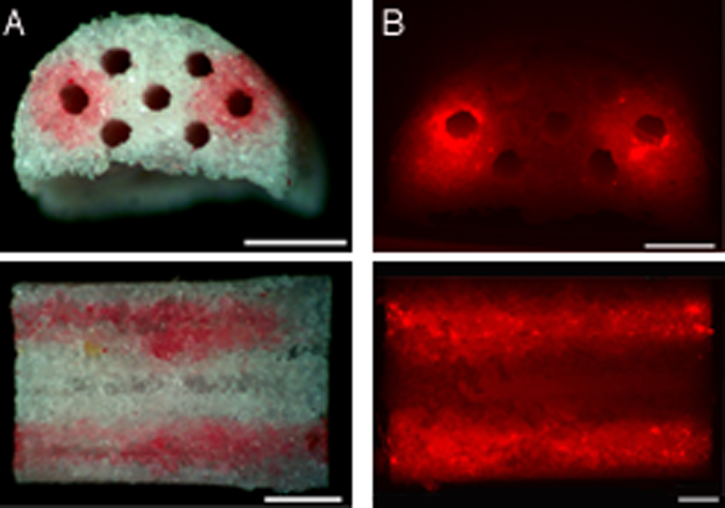
Figure 1.

Patterning of fibronectin/lipoplex mixture within the outer two channels of the middle row of the multiple channel bridge. Bridges were washed twice after deposition. A) Sirius red stain of fibronectin inside the deposition channels. From top to bottom: side view and cross section of bridge. Scale bars: 1 mm. B) Fluorescence image of rhodamine labeled DNA (red), complexed with Transfast and mixed with fibronectin inside deposition channels. From top to bottom: side view and cross section of bridge, Scale bars: 500 µm.
