
Figure 5.
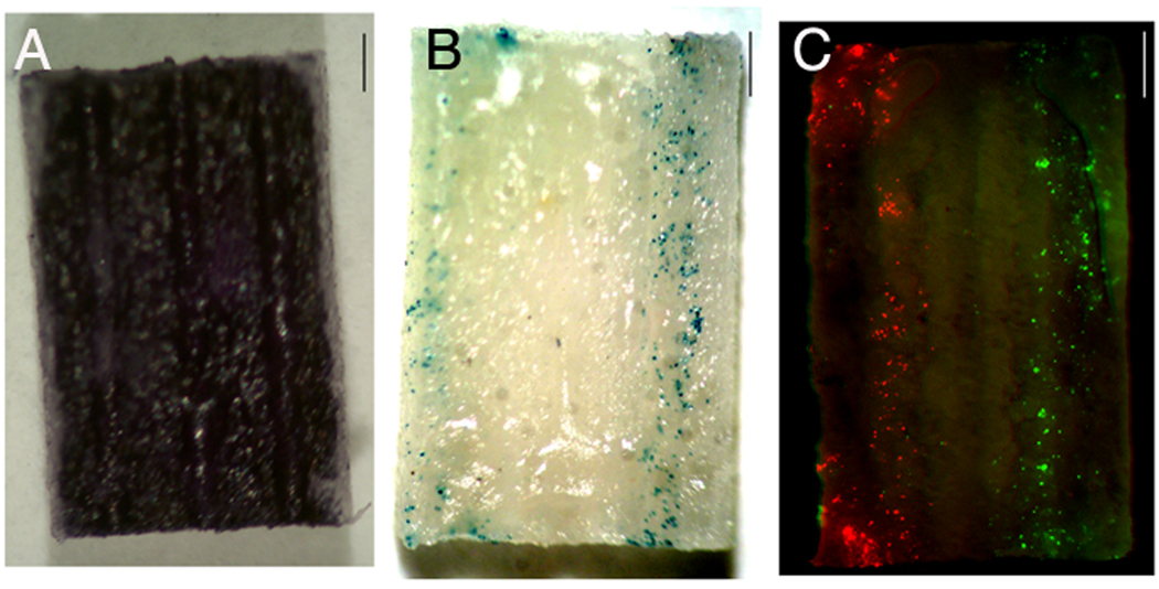
Patterned transgene expression in channels of 3D multiple channel bridges. A) MTT stain for metabolically active cells, 48 h after cell seeding (dark blue). B) Transfected cells (blue) 48 h after cell seeding, with pβgal deposited only in the two outer channels of the middle row of the bridge. C) Transfected cells (green respectively red), for 2 plasmids, pGFP and pDsRed, deposited in the two outer channels of the middle row. Scale bars: 1 mm.