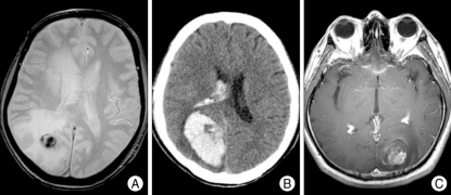
Fig. 1.
A : Gradient echo magnetic resonance (MR) image showing a hemorrhagic component in the tumor as lesions with low signal intensities. B : A large intracerebral hemorrhage (ICH) is identified on computed tomography scan of a patient with sudden headache and visual disturbance. The area near the midline in the hematoma with lower density than hematoma is brain metastasis from hepatocellular carcinoma. C : An ICH and edema around the heterogeneously enhancing mass on T1 gadolinium-enhance MR image.

