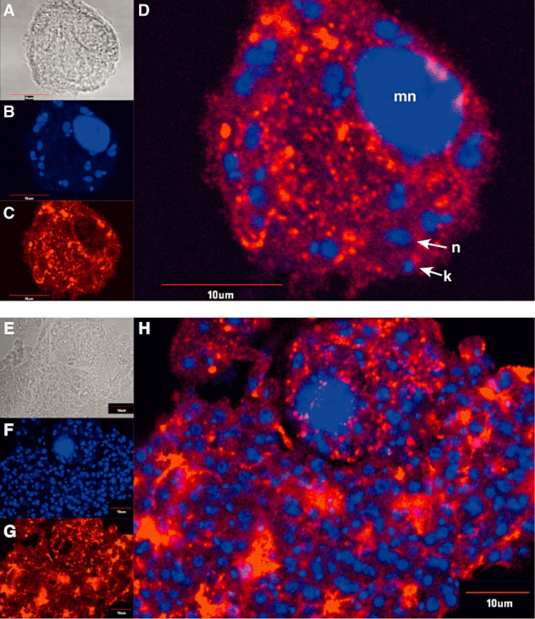
Fig. 6.

P46 localization in infected macrophages. Bone marrow-derived macrophages were infected with L. major (A–D) or L. major[pcosP46] (E–H) at a 1:10 cell ratio and stained with anti-P46 antiserum and DAPI. Bright field images (A,E), DAPI stain (B,F) and anti-P46 stain (C,G) are shown. (D,H): DAPI and anti-P46 overlays. mn = macrophage nucleus; k = kinetoplast; n = parasite nucleus. 10 µm size bars are shown.
