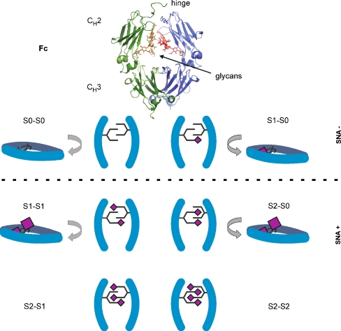
Fig. 1.
Sialylation of the Fc fragment. The Fc fragment of an antibody (taken with Pymol from 1HZH.PDB; top picture). The CH2 domain in all conceivable glycoforms in frontal view and—except for the hypersialylated glycoforms—in side view (bottom picture). In the last row, only a second sialic acid (purple diamond) is exposed and accessible to a lectin

