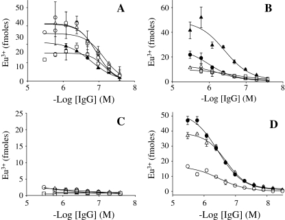
Fig. 1.
Aβ40 conformer binding by IgGs contained in normal plasma. Representative antibody binding curves for normal plasma IgGs (filled circle, unfilled circle, unfilled square, filled triangle, unfilled triangle) against Aβ fibrils (a), CAPS (b), and Aβ monomers (c). d Binding curves for high (filled circle) and low (unfilled circle) reactive plasma pools, and for an equimolar mix (unfilled triangle) against Aβ fibrils. Each plasma pool consisted of 15 plasma specimens. CAPS indicates dityrosine cross-linked β-amyloid protein species

