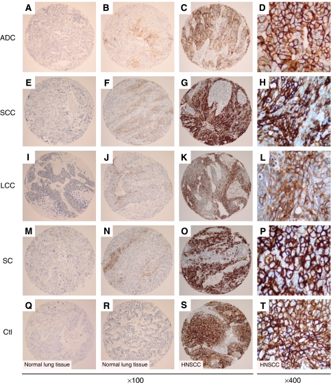
Figure 1.
CAIX expression detected by immunohistochemistry in tissue microarray cores. Staining levels for CAIX in NSCLC histological subtypes: low (A, E, I, M), intermediate (B, F, J, N) and strong (C, G, K, O). CAIX membrane staining in adenocarcinoma (ADC) (A–D), squamous cell carcinoma (SCC) (E–H), large cell carcinoma (LCC) (I–L), and sarcomatoid carcinoma (SC) (M–P). Normal bronchial epithelium (Q) and alveolar tissue (R) are devoid of staining. Strong membrane staining in head and neck squamous cell carcinoma (HNSCC) cores used as positive control of CAIX immunostaining (S–T). Panels D, H, L, P and T are higher magnifications showing details of cells within the corresponding tumour shown on panels C, G, K, O, and S, respectively.

