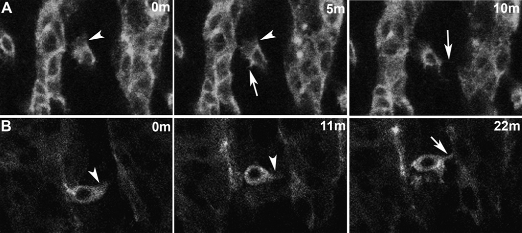
Figure 3. Dynamic actin-bases behaviors of myoblasts.
Lateral views of live twi promoter-GFP::actin embryos. Actin labeling is concentrated at the cell cortices and in cellular extensions such as lamellipodia and filopodia. Such behaviors appear critical for myoblast fusion. The nucleus is evident as a non-labeled structure in the middle of the cells. Each panel represents a single time point from a time lapse sequence. Images are single optical slices.
(A) FC for a segment border muscle prior to fusion extends lamellipodia (arrowhead) and filopoida (arrow). (B) FCM extends lamellipodia (arrowhead) and a filopodium (arrow) prior to fusion.

