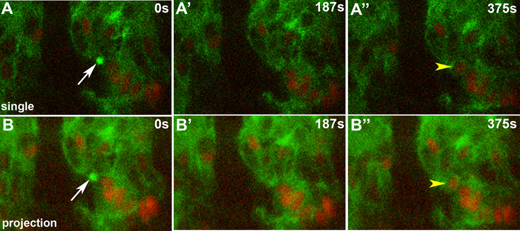
Figure 4. Single myoblast fusion event.
Lateral view of live twist promoter-GFP::actin, apME580-NLS::dsRed.T4 embryo. Each row of panels represents a time point from a time-lapse sequence. (A) Single optical slice. In this sequence, an actin focus (white arrow) forms at the site of adhesion between an FCM and an apterous-labeled myotube. This focus resolves, followed by fusion and addition of an additional labeled nucleus (yellow arrowhead) to the myotube. (B) Same sequence as in A, but as an optical projection displaying 9 µm of the Z-axis.

