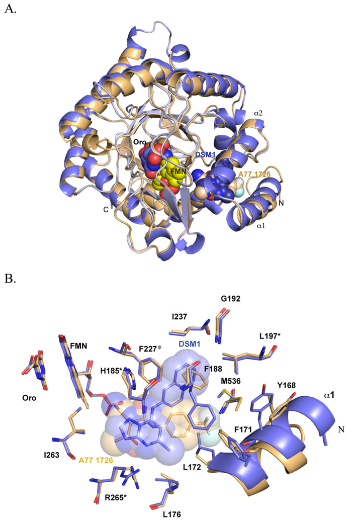
Fig. 3.
X-ray structure of PfDHODH. A. Ribbon diagram of an alignment of the structures bound to A77 1726 (tan; pdb 1TV5) and DSM1 (purple; pdb 3I65). B. A77 1726 and DSM1 inhibitor binding sites. Residues marked with * are conserved in human DHODH, while the remainder of the shown residues are variable between Plasmodium and human DHODH. Structures were displayed in PyMol (Delano Scientific)

