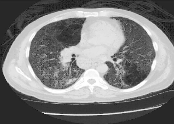
Figure 4.

Patient 5. HRCT scan at the level of the left atrium demonstrating diffuse ground glass attenuation with fine reticulation and cystic changes. Lymph node enlargement involving the azygoesophageal recess is seen. Clinical characteristics of the patient are shown in Table 4
