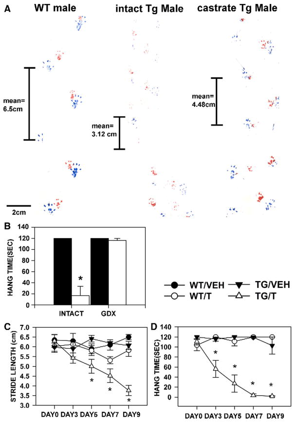
Fig. 2.
Androgen dependence of motor dysfunction in HSA-AR Tg mice. Tg males exhibit impairments in motor tasks such as gait analysis and Hang test, which are somewhat normalized by castration. A) The HSA-AR male’s paw prints (blue for rear paws and red for front paws) are shown prior to castration (middle) and following castration (right). A WT brother’s prints are shown for comparison (left). Stride length, which is related to strength, for each mouse is indicated. B) Latency to fall when held suspended from a cage lid (hang time) is significantly shorter for HSA-AR Tg males (open bars) relative to WT brothers (filled bars) prior to castration (INTACT), but not following castration (CASTRATE). Androgen dependence is also indicated by androgen administration to HSA-AR Tg females (C, D). In this experiment, HSA-AR Tg females (triangles) are compared to WT sisters (circles), all animals are given either testosterone in a silastic capsule (T — empty symbols) or the vehicle capsule alone (VEH — closed symbols). Motor dysfunction is only observed in T treated Tg females (TG/T —open triangles) and begins 3 days following T treatment. Graphs represent mean ± SEM and * indicates significantly different from VEH as indicated by PLSD comparisons with p<0.05.

