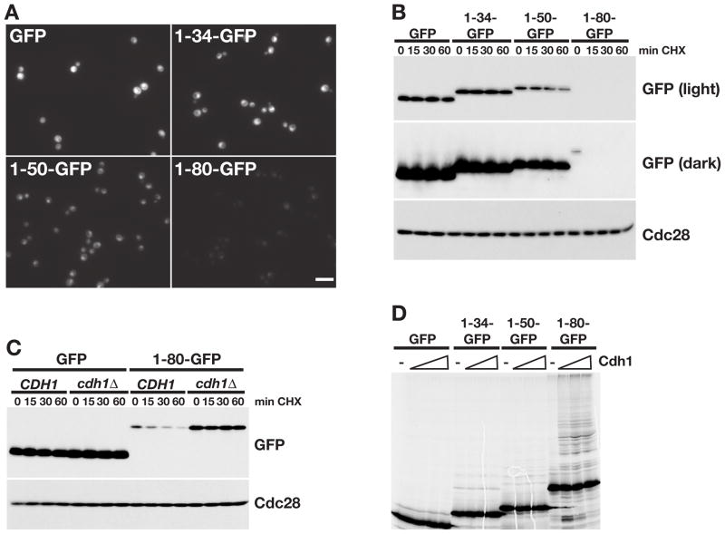
Figure 5. The N-terminus of Cik1L is an APC/C degron.
(A) GFP expression in cells expressing GFP or Cik1-GFP fusions (1-34-GFP, 1-50-GFP, 1-80-GFP) from the TEF promoter. Scale bar represents 10μm. (B) Cycloheximide chase assay of the indicated GFP proteins. Cells from (A) were arrested with α factor and treated with cycloheximide for the indicated number of minutes (min CHX). Proteins were analyzed by GFP Western blot (light and dark exposures are shown). Cdc28 is shown as a loading control. Cell cycle profiles are shown in Figure S5A. (C) Cycloheximide-chase assay of GFP and 1-80-GFP in asynchronous CDH1 and cdh1Δ cells. Cell cycle profiles are shown in Figure S5B. (D) GFP and Cik1-GFP fusion proteins were translated in vitro and equivalent amounts were incubated with purified E1, E2, ATP, ubiquitin, and APC/C in the absence (−) or presence of Cdh1. Reactions were analyzed by SDS-PAGE and visualized with a phosphorimager, quantitation is shown in Figure S5C.

