
Figure 5.
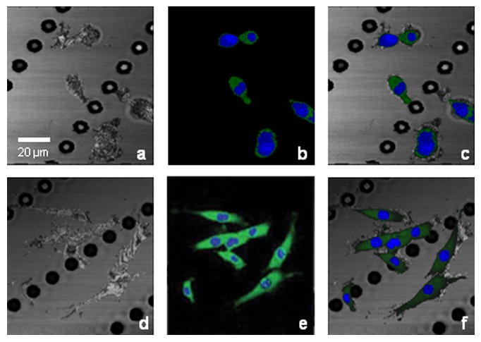
Integrated OCM/MPM imaging of GFP-vinculin fibroblasts seeded on a microtextured substrate under static (a-c) and dynamic (d-f) culture. (a) and (d) OCM images showing both cells and substrate. (b) and (e) MPM images showing nuclei (blue channel) and GFP-vinculin (green channel) denoting cell-cell and cell-substrate adhesions. (c) and (f) Multimodal images of combined OCM/MPM showing structural and functional relationships between cells and substrate/scaffold. The white arrow indicates the direction of stretching. Scale bar applies to all images.