
Fig. 6.
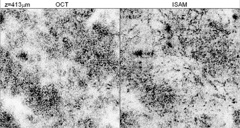
Human breast tumor tissue imaged with OCT (left) and with the real-time 2D ISAM system (right), and post-processed in slow scan direction. The en face slice shown in the plane of the page is 450 μm above the focus. The volumetric ISAM data shows cell and nuclei data not observed in the volumetric OCT data. The scale bar is 100 μm. A portion of the real-time 2D ISAM acquisition (movie2.avi, 6.1 MB) and a representative en face fly-through (movie3.avi, 15 MB) are available online.