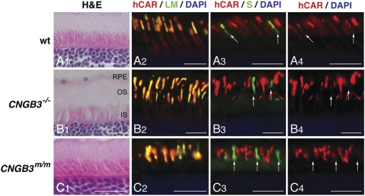
Figure 2.
Presence of L/M- and S-cones in CNGB3-mutant canine retinas, expressing markers that characterize the differentiated state. Comparison of canine retinal structure and cone-specific protein expression in normal (A1—A4; 12 weeks), CNGB3−/− (B1—B4; 17 weeks), and CNGB3m/m (C1—C4; 12 weeks) dogs. The H&E-stained sections show normal outer retinal structure independent of disease status (A1, B1, C1). The expression and distribution of L/M- and S-opsin is not affected by the CNGB3-mutation. L/M-opsin labeling (green) co-localizes with cone arrestin expression (hCAR labeling in red) in the outer segments of the L/M-cones (A2, B2, C2). In contrast, cone arrestin labeling is weak in S-cone outer segments; hence, the co-localized signal is dominated by the green S-opsin labeling (arrows in A3, B3, and C3). Closer analysis of cone arrestin distribution shows that the hCAR labeling is much weaker in the S-cone outer segments compared with the L/M-cones of the canine WT retina (arrows in A4). hCAR labeling is essentially not visible in the S-cone outer segments of the CNGB3-mutant dogs (arrows in B4 and C4). Calibration bars = 20 µm. RPE, retinal pigment epithelium; OS, outer segment; IS, inner segment.

