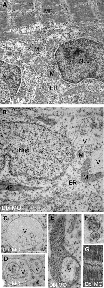
Figure 8.
Ultrastructural analysis of MTMR14/MTM1 double-morphant skeletal muscle. Electron micrographs of skeletal muscle from 48 hpf embryos. (A and B) Low magnification of MTM1/MTMR14 double morphants (Dbl MO) revealed a paucity of sarcoplasm, decreased endoplasmic reticulum density (ER) and the presence of abnormal vacuolar structures (V) and abnormally appearing mitochondria (M). (C–E) Higher magnification examination of the abnormal vacuolar structures, including a vacuole within a mitochondrion (E) and vacuoles with subcellular contents (F). (G) Despite these changes, myofibrils appeared normal. Scale bars = 500 nm.

