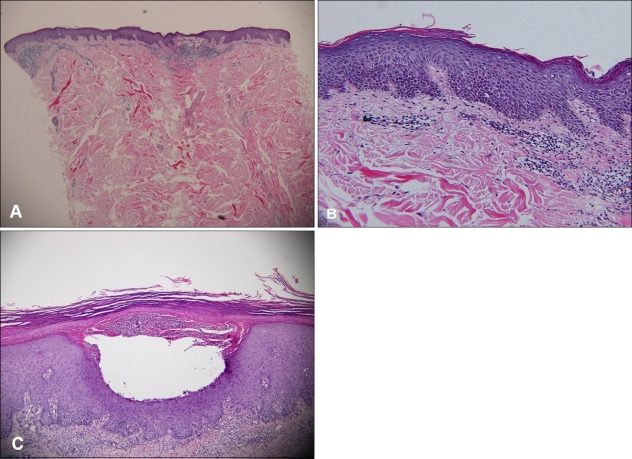
Fig. 2.
Epidermal hyperplasia with hyperkeratosis and parakeratosis are shown on horny layer; granular layer has disappeared (A, B). Capillaries in the papillary dermis associated with perivascular lymphocytic infiltration (B). Munro microabscess was shown. Intraepidermal pustule formation was shown (C) (H&E, A: ×40, B: ×100, C: ×200).

