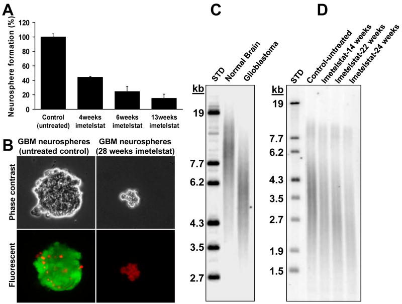
Figure 3. Long-term treatment with imetelstat in vitro leads to decrease clonogenic capacity, telomere shortening and cell death.
A. Clonogenic neurosphere formation assay of GBM tumor-initiating cells exposed to imetelstat. A total of 500 cells were plated on 10cm dishes, fresh media was exchanged after 5 days and the neurospheres were counted after 10-14 days; B. Live/dead assay on the long term-treated cells versus the untreated controls. Green cells - Calcein AM (live cells), red cells - ethidium homodimer (necrotic cells); C. TRF gel shows that GBM tumor cells have short telomeres (average about 6kb) compared to normal brain (∼10-12kb); D. TRF analysis of another primary glioma that has even shorter initial average telomere length (∼3.5-4kb) indicates progressive telomere shortening in GBM tumor-initiating cells treated in vitro 2×/week with 2μM imetelstat.

