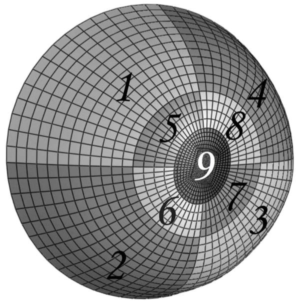
Figure 2.
Anatomically accurate geometry of one posterior scleral shell (from the clamping boundary to the ONH) that was reconstructed from experimental topography and thickness measurements. Regions 1–4 encompass the peripheral sclera, Regions 5–8 the peripapillary sclera and Region 9 the ONH. The peripapillary sclera (Regions 5–8) extended approximately 1.5 to 1.7 mm from the scleral canal, which was defined as the border between the ONH (Region 9) and the peripapillary sclera (Regions 5–8). The clamping ring was located approximately 3 mm posterior to the equator.

