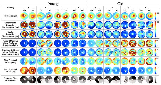
Figure 5.
Individual results for all posterior scleral shells as viewed from the back of the eye (Superior is up). Scleral thickness was experimentally measured at IOP = 5 mmHg and interpolated to obtain continuous thickness maps. Tangent modulus, structural stiffness, maximum principal stress and strain are shown for all eyes at a single IOP of 30 mmHg. Good agreement is observed between FE-computed and experimentally measured posterior displacements (plotted for an IOP range of 5–30 mmHg). Finally the preferred fiber orientation is shown for all eight regions of each eye, where // (black) corresponds to a collagen fiber organization tangent to the scleral canal (circumferential, θp = 0°) and ⊥ (white, θp = 90°) corresponds to a fiber organization that is perpendicular to the scleral canal (meridional). Note that the data for the two eyes of each monkey are much more similar than between monkeys, and there are clear age-related differences in all measures except for preferred collagen fiber orientation.

