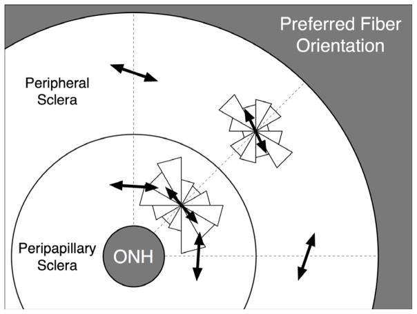
Figure 6.
Pooled distributions (all eyes) of the preferred fiber orientation in both the peripapillary and peripheral scleral regions are shown as two symmetric rose diagrams, in which larger triangles indicate the most commonly derived orientations. Mean preferred fiber orientations (black arrows) were computed for each of both distributions using a circular statistics formula56 and were equal to 176.2° and 162.0° in the peripapillary and peripheral sclera, respectively. Note that 0° or 180° correspond to an orientation tangent to the scleral canal and 90° to an orientation perpendicular to the scleral canal. On average, this result suggests a tendency toward a circumferential organization of the collagen fibers around the scleral canal in both the peripapillary and the peripheral sclera.

