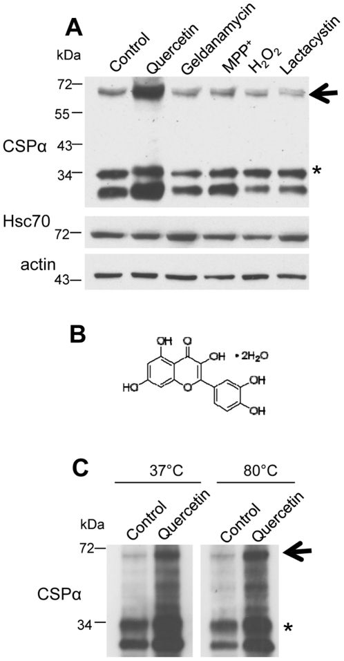
Figure 1. Quercetin promotes CSPα dimerization.
(A) CAD cells were transiently transfected with 0.5 µg of c-myc-CSPα DNA and treated with the indicated agent (200 µM quercetin, 1 µM geldanamycin, 1.5 µM MPP+, 0.2 mM H2O2 or 10 µM lactacystin) for 24 hours prior to lysis. 40 µg of cellular protein was resolved by SDS-PAGE and CSPα and Hsc70 were detected by Western analysis. β-actin is shown as a loading control. (B) Chemical structure of quercetin dihydrate. (C) CAD cells were transfected with 1.0 µg of c-myc-CSPα DNA and treated with 100 µM quercetin for 24 hours prior to lysis. 30 µg of protein was heated at either 37°C or 80°C for 10 minutes prior to being resolved on an SDS-PAGE gel. CSPα was detected by Western analysis with a c-myc antibody. Arrows indicate CSPα dimer at ∼72 kDa; asterisks indicates palmitoylated CSPα monomer at ∼34 kDa. Data are representative of three separate experiments.

