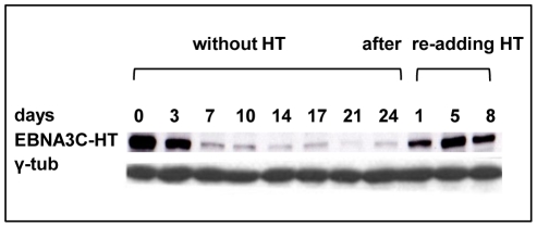
Figure 1. EBNA3C inactivation and reactivation in LCL 3CHT.
Cells that had been grown in culture medium containing HT were washed and re-suspended in medium with HT omitted. After culturing for the number of days indicated, protein extracts were western blotted and probed with an anti-EBNA3C MAb to reveal EBNA3C-HT. After 17 days some cells were transferred to medium containing HT and after 1, 5 or 8 days samples were again taken for western blotting. The western blot was re-probed with anti-γ-tubulin to ensure equal loading of the proteins.

