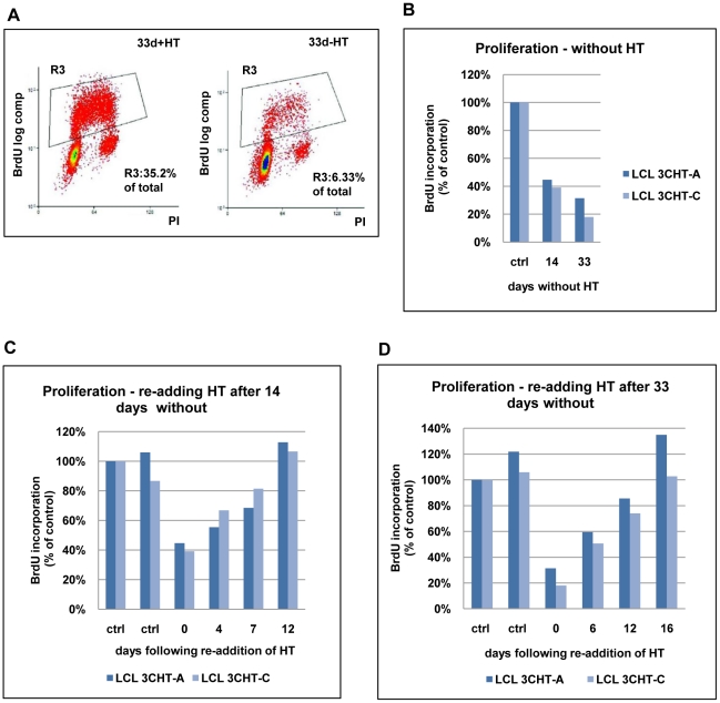
Figure 2. Proliferation of LCL 3CHT after inactivation and reactivation of EBNA3C.
(A) LCL 3CHT cells were cultured either with HT (+HT) or for 33 days after the removal of HT (-HT) from the growth medium. Cells were pulsed for 1 hour with BrdU, harvested, fixed and stained with anti-BrdU-FITC and propidium iodide. The cells were analysed by flow cytometry. The gated BrdU-positive population is reduced in the absence of HT. (B) Two LCL 3CHT (-A and -C) generated from independent 3CHT-BACs using PBL from a single donor were analysed as in (A) after 14 and 33 days without HT. Histograms show BrdU incorporation relative to the control, cycling population. The control (ctrl) was a proliferating population of LCL 3CHT grown in medium with HT. (C) LCLs 3CHT-A and -C were cultured for 14 days in the absence of HT (0) and BrdU-incorporation was assayed as in (A). The incorporation of BrdU was also assayed as above after 4, 7 and 12 days after re-adding HT. (D) A similar experiment was performed on cells cultured without HT for 33 days.

