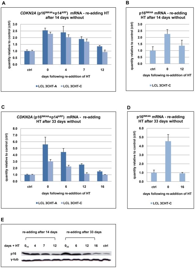
Figure 3. Repression of p16INK4A following reactivation of EBNA3C.
(A) After 14 days without HT (0), total RNA was extracted from aliquots from two LCL 3CHT populations (-A and -C). HT was re-added to the remaining cells and further RNA samples were taken at the times indicated. Real time quantitative RT-PCR (qRT-PCR) was performed to quantify CDKN2A transcripts. The histogram corresponds to CDKN2A mRNA relative to that in control cycling populations of each LCL 3CHT. (B) As in (A) but using a p16INK4A-specific qPCR. (C) & (D) Similar assays to those described in (A) and (B) after 33 days without HT. (E) Western blots – probed with a p16INK4A-specific MAb – of protein extracts from LCL 3CHT-C cells to which HT was re-added after 14 or 33 days in its absence (014 and 033 respectively). The control (ctrl) is LCL 3CHT-C cells continuously cultured in medium with HT.

