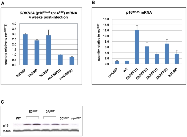
Figure 9. Expression of p16INK4A is increased in CtBPM LCLs relative to revertant and WT LCLs.
(A) E3CtBP, 3ACtBP, 3CCtBP and two revCtBP LCLs were harvested 28 days after the infection of primary B cells with the recombinant EBVs. RNA was extracted and the relative levels of CDKN2A transcripts were quantified by qRT-PCR. (B) RNA was extracted from two established E3CtBP LCLs, two 3ACtBP LCLs, 3CCtBP LCL, a revertant (revCtBP) and a WT-EBV LCL. The relative levels of p16INK4A transcripts were quantified by qRT-PCR. (C) Western blot analysis of protein extracts from two E3CtBP LCLs, two 3ACtBP LCLs and a 3CCtBP LCL all established from a single donor. A revertant (revCtBP) and a WT-EBV LCL are shown for comparison. Levels of p16INK4A (p16) are shown relative to γ-tubulin (γ-tub).

